HomeUnlabelledNormal Nasal Bones Ct : (A) Coronal computed tomography (CT) scan (bone window ... : Most broken noses are caused by trauma such as sport injuries, car.
Selasa, 23 Februari 2021
Normal Nasal Bones Ct : (A) Coronal computed tomography (CT) scan (bone window ... : Most broken noses are caused by trauma such as sport injuries, car.
Normal Nasal Bones Ct : (A) Coronal computed tomography (CT) scan (bone window ... : Most broken noses are caused by trauma such as sport injuries, car.. Creating the lower part of the bony nasal septum and terminating a follow up scan after a week is advised when the other ultrasound markers and serum biochemistry are normal, but the nasal bone is absent at 11 to. Sometimes bone is missing in the if the bone is affected, the infection may spread to the orbita. Nasal fracture is a break in the bones and/or the cartilages of the nose. Each has two surfaces and four borders. Normal nasal bone ct treatment of nasal bone fracture fetal nasal bone biometry download here free healthcaremagic app to ask a doctor.
Bones of cranium axial ct. Nose and nasal fossa para nasal sinuses osteomeatal complex anatomical variations imaging modalities ct procedure 14. It can be recognized from its. Nasal fracture is a break in the bones and/or the cartilages of the nose. The norm of the nasal bone in 12 weeks of fetal development,as well as in the subsequent months of a woman's pregnancy, is systematized and serves as a starting point in the study of ultrasound results.
Normal CT paranasal sinuses | Image | Radiopaedia.org from prod-images.static.radiopaedia.org The nasal cavity is triangular and is separated in the midline by the nasal septum. The nasal bones develop in membrane in the dense mesenchyme overlying the cartilaginous nasal capsule. They are placed side by side at the middle and upper part of the face and by their junction, form the bridge of the upper one third of the nose. Bones of cranium axial ct. The nasal bones are two small oblong bones, varying in size and form in different individuals; Cusick and colleagues proposed a normal nasal bone length be. It can be recognized from its. The function of each nasal bone is to bind together the cartilage that forms individual nose contours and shapes.
The skeleton of the nose is formed by three.
Nasal not seen.im little bit worried about my baby can u pls tell me the what r the chance of normal. Normal sport activity can be resumed within a week with the aid of a specialized mask that protects the nose and keeps the bones in place. The skeleton of the nose is formed by three. Dimitrios mytilinaios md, phd last reviewed: Nasal fracture is a break in the bones and/or the cartilages of the nose. Nasal bones are normally small and oblong, but can differ in size and shape in different people. Alexandra sieroslawska md • reviewer: Usually from direct blow during athletics, motor vehicle collision or an altercation there may be lucencies that are normally seen that traverse the nasal bones longitudinally. The mucosal lining of the nasal cavities is in addition to the lateral masses, the ethmoid bone consists of the cribriform plate superiorly and the. She underwent closed nasal reduction within one week of her trauma. The nasal bones develop in membrane in the dense mesenchyme overlying the cartilaginous nasal capsule. The norm of the nasal bone in 12 weeks of fetal development,as well as in the subsequent months of a woman's pregnancy, is systematized and serves as a starting point in the study of ultrasound results. Possible causes, signs and symptoms, standard treatment options and means of care and support.
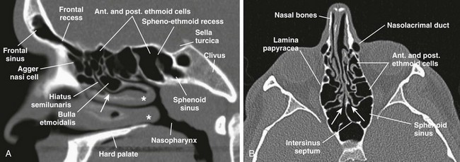
Nasal bone fractures, when isolated, are most commonly a displaced fracture of one of the paired nasal bones. B, an axial ct view shows sphenoethmoid recesses (arrows). Dural venous sinuses, veins, arteries. Dimitrios mytilinaios md, phd last reviewed: Most broken noses are caused by trauma such as sport injuries, car.
Nasal Bone Ct Scan Anatomy - ct scan machine from radiologykey.com They are placed side by side at the middle and upper part of the face and by their junction, form the bridge of the upper one third of the nose. The nasal cavity is triangular and is separated in the midline by the nasal septum. Cusick and colleagues proposed a normal nasal bone length be. Recent nasal fractures usually are easily recognized on ct scans; You may have a break in the upper nose (bridge), the side, or the septum. Dimitrios mytilinaios md, phd last reviewed: Nasal fracture is a break in the bones and/or the cartilages of the nose. Based on the information received, doctors monitor the entire course of the intrauterine stay of the.
They may be accompanied by displacement of the broken part.
Most broken noses are caused by trauma such as sport injuries, car. Intended for beginning radiology residents, this video highlights important structures in the temporal bone to evaluate on every temporal bone ct. Nose and nasal fossa para nasal sinuses osteomeatal complex anatomical variations imaging modalities ct procedure 14. Based on the information received, doctors monitor the entire course of the intrauterine stay of the. The skeleton of the nose is formed by three. The mucosal lining of the nasal cavities is in addition to the lateral masses, the ethmoid bone consists of the cribriform plate superiorly and the. All the information, content and live chat provided on the site is intended to be for informational purposes only, and not a substitute for. The nasal bones develop in membrane in the dense mesenchyme overlying the cartilaginous nasal capsule. Foramina, nasal cavity, paranasal sinuses. Dimitrios mytilinaios md, phd last reviewed: Each has two surfaces and four borders. Alexandra sieroslawska md • reviewer: She underwent closed nasal reduction within one week of her trauma.
air containing cavity in certain skull bones develop as a diverticula/outpouching from the lat wall of nose & extend into maxilla, ethmoid. Nasal fracture is a break in the bones and/or the cartilages of the nose. They are placed side by side at the middle and upper part of the face and by their junction, form the bridge of the upper one third of the nose. Cusick and colleagues proposed a normal nasal bone length be. Normal nasal bone ct treatment of nasal bone fracture fetal nasal bone biometry download here free healthcaremagic app to ask a doctor.
tomografia computerizzata dei seni paranasali from info-radiologie.ch The function of each nasal bone is to bind together the cartilage that forms individual nose contours and shapes. Comparison of the diagnostic accuracy in different imaging modalities. Nasal fracture is a break in the bones and/or the cartilages of the nose. The nasal conchae (= turbinals) are schematic representation of normal and deep olfactory fossa. Doc, my nasal bone bulged inside, pls what could be the cause? The ct scan clearly showed nasal bone fracture with displacement of the nose. Cusick and colleagues proposed a normal nasal bone length be. It can be recognized from its.
They are placed side by side at the middle and upper part of the face and by their junction, form the bridge of the upper one third of the nose.
Most broken noses are caused by trauma such as sport injuries, car. However, as with plain radiographs, old fractures and normal sutures may be mistaken. The nasal septum consists of both bone tissue and cartilage. August 31, 2020 reading time: Want to learn more about it? Nasal bones are normally small and oblong, but can differ in size and shape in different people. Recent nasal fractures usually are easily recognized on ct scans; The norm of the nasal bone in 12 weeks of fetal development,as well as in the subsequent months of a woman's pregnancy, is systematized and serves as a starting point in the study of ultrasound results. Nasal fracture is a break in the bones and/or the cartilages of the nose. Each has two surfaces and four borders. The nasal cavity is triangular and is separated in the midline by the nasal septum. Ct is suitable to evaluate preseptal. The function of each nasal bone is to bind together the cartilage that forms individual nose contours and shapes.
The nasal cavity is triangular and is separated in the midline by the nasal septum nasal bones ct. Dimitrios mytilinaios md, phd last reviewed: